M700 Automated Perimeter
Value. Proven. Reliable.
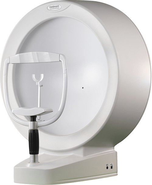
Perimetry you can count on
The Medmont M700 leverages over 30 years of proven perimetry technology. It’s a reliable, effective and economical tool for assessing visual fields.
Built to last
Recognised as a value leader for its reliable performance and longevity, the hand assembled M700 is built to last.

Reliable and durable
The fully automated unit, with no moving parts and standard computer hardware, resulting in minimal maintenance requirements. There are no routine service requirements, and we see units in use for over 20 years.

Effective and efficient
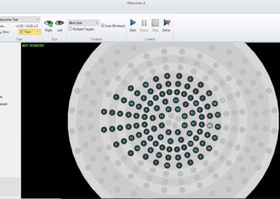
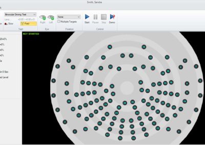
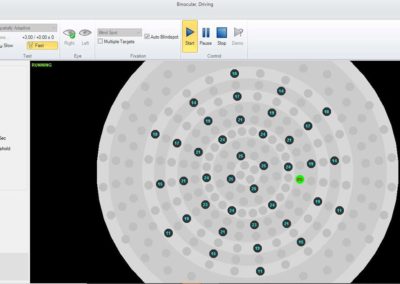
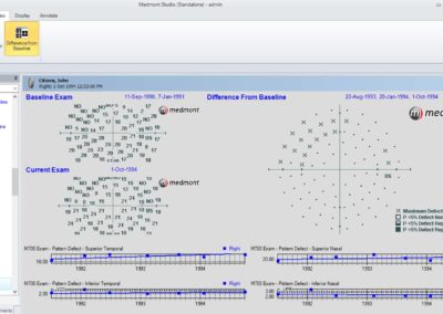
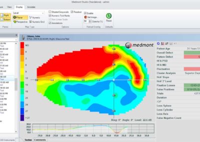
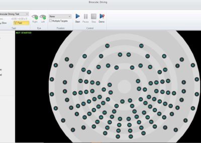
The M700 uses a radial pattern that provides a high density of test points in central regions. It’s designed to be comparable but more sensitive to glaucoma than standard 24-2 and 30-2 tests. With the M700, you can add points and retest during an exam, saving precious time that comes with rework.

Personalised patient care
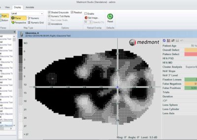
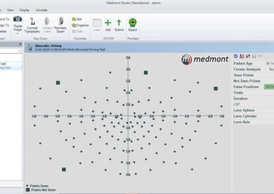
The M700 uses standard static perimetry to perform rapid and reliable screening and visual field threshold tests to assess visual field loss relating to glaucoma, macula, neurological, peripheral and binocular vision, etc. Its standard suite of protocols meets diverse patient needs, and the custom template builder lets you personalise care.
Media Gallery
M700 Key Features
Full Field Coverage (160°) |
Advanced Fast Threshold Testing Strategy, Employing Baysean Testing Techniques |
Flicker Test Facility, With Proven Early Field Loss Detection Capabilities |
Patient Reliability Indicators • False Positives |
Field Analysis Tools • Glaucoma Progression |
Display Options • Grey Scale |
Result Outputs • Customizable Page Layout |
Regression Graphs Printing • Pattern Defect Index |
Specifications
Size
- Stimulator Unit Dimensions:
- 626mm Wide x 438mm Deep x 713mm High
- Stimulator Unit Weight:
- 12kg
- Shipping Dimensions / Weight:
- 71cm x 52cm x 85cm, 23 kg (Unit and Box)
Performance
- Stimulator Screen:
- Part Hemispherical Bowl Radius 30cm Integrated Diffusing Surface
- Test Fields:
- Binocular 30°/40° : 21-128 Points Binocular Driving 80° : 119 Points Central 22A 22° : 45 to 96 Points Central 30° : 100 Points Driving Test 50°/80° : 103 Points Flicker 15°/22° : 69 Points Full 50° : 164 Points Glaucoma 22°/50° : 104 Points Macula 10° : 49 Points Neurological 50° : 164 Points Peripheral 30° to 50° : 73 Points Flash Scan 22°/30° : 40 Points Spatially Adaptive 50° : 39 to 168 Points CV% 100 Points 60° : 100 Points Central 22 22° : 50 Points
- Stimulus Source:
- Rear Projection Light Emitting Diode
- Stimulus Color:
- Pale Green 565nm Half Bandwidth 28nm Red 626nm (Optional)
- Stimulus Size:
- Goldmann Size III (0.43°), Model CR Red (0.72°) (Optional)
- Stimulus Intensity:
- 16x3dB Levels 0db (Max Brightness) to 45dB (Min Brightness) +/-1dB
- Stimulus Duration:
- Adjustable: 0.1 to 9.9 sec. (nom. 0.2 sec)
- Patient Response Time:
- Adaptive to Patient Speed, Operator Selection of Normal or Slow Ranges Adjustable: 0.1 to 9.9 sec (nom. 1.1 sec)
- Minimum Inter-stimulus Delay:
- Adjustable 0.1 to 9.9 sec (nom. 0.4 sec)
- Background Illumination:
- 10 asb (3.2cd/m2), Automatic Level Control 31.5 asb (10cd/m2) – German Driving Test
- Test Lens Diameter:
- 38mm
- Fixation Method:
- Automatic Iris Tracking During Test with Relaxed Tolerance. Heijl-Krakau Blind Spot Method. Visual and Audible Warning of Fixation Errors. Video Fixation Monitoring
Electrical
- Stimulator Unit Power:
- 100-240 VAC 50-60Hz , 0.25-0.15A
Computer Minimum Requirements
- PC and Mains Powered Peripherals: EN/IEC60950 Compliant
- Supported PC Operating Systems (OS’s):
- Windows 11 Professional and Home (64 bit) Professional.
- Windows 10 Professional and Home Professional (64 and 32 bit).
- Windows Server 2016, 2019 and 2022 (64-bit editions only).
- Windows Server “Server Core” installations are not supported.
- Processor: Intel™ Generation 6 i5 or later
- Motherboard: Genuine Intel™ chipset highly recommended
- VIA chipsets have proven to be unreliable
- Memory: 8 GB for non-video captures, 16 GB for video captures.
- Hard Disk Space: 40 GB for non-video, 200GB for video captures (the more the better)
- Video cards: GPUs with dedicated memory of 2 GB or more are recommended. GPUs that share memory require 128 MB of memory minimum and are not recommended.
- Screen resolution: 1920 x 1080 recommended, minimum supported 1280 x 720.
- Printer: Bubble-jet or Laser-jet printer (Color / Monochrome).
- USB: At least 1 USB 2.0 Compliant port on the PC.
ALWAYS FOLLOW THE DIRECTIONS FOR USE
Brochure
Request a free virtual demo

