medmont meridia™ Advanced Topographer
Expand patient outcomes and practice profitability.

Set your practice apart with the gold-standard of specialty lens care.
Our next-generation topographer takes corneal mapping to the next level. Quickly embraced by contact lens specialists worldwide, the Medmont Meridia™ builds on the trusted E300 with full-colour maps, an ultra-wide field of view, and unparalleled corneal coverage. And that’s just the start.
The Meridia™ Pro adds a range of new imaging modes for the anterior eye — including dry eye (DED) analysis and reporting, tear film assessment, meibography, and more. And the all-new Meridia™ Vantage rounds out the range with scleral topography, refined ergonomics and a bold, modern design. It’s everything you need for patient care and practice success, all packed into one compact, reliable, easy-to-use instrument.
Request a free virtual demo
See clearly, treat optimally
Access comprehensive suite of high-resolution images, a wider field of view, and extra applications to boost your practice’s ability to care for patients.

Lift your care to a new standard
The Medmont Meridia™ builds on the E300’s 20-year legacy, delivering reliable, best-in-class accuracy for contact lens fitting — with powerful new features designed to elevate patient care and maximise your practice’s ROI.
With a larger field of view, the Meridia captures more of the patient’s eye in a single full-colour image. Combined with Medmont’s trusted composite map technology, it gives you clearer, more confident evaluations and fittings—leading to better first-fit success and reduced chair time.
The Meridia offers a versatile, all-in-one platform for your practice. Alongside advanced corneal topography, it includes premium anterior and fluorescein imaging and video, enhanced meibography, clear tear meniscus height and tear break-up time measurements, and more. This new standard in Medmont topography helps you see more patients efficiently while expanding your dry eye and specialty lens services — all while improving your bottom line.

Patient engagement
The Meridia is designed to boost patient engagement, helping improve compliance, encourage repeat visits, and generate more referral business. Paired with Medmont Studio, it offers universal grading scales and visual reports that make consultations and treatment planning straightforward and effective.

Ergonomic design
The Medmont Meridia platform features an advanced ergonomic design with five quick keys on the instrument. Combined with intuitive software guidance, this all-in-one system supports efficient workflows and optimises space — making it a standardised solution you can rely on.
Media Gallery
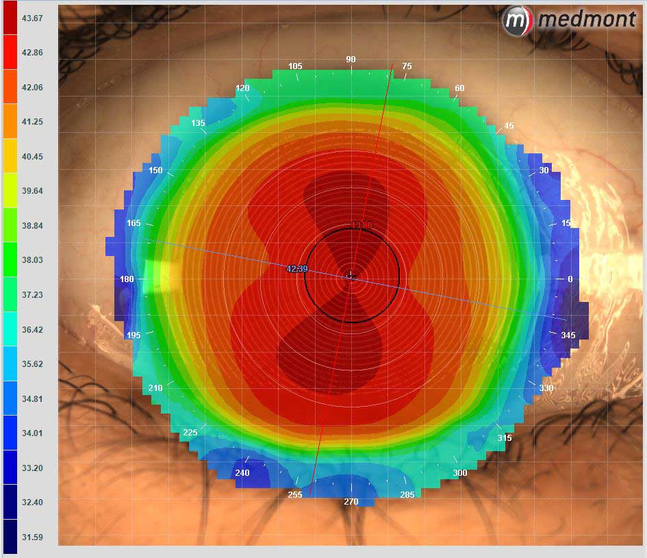
WTR Astigmat Composite Topo
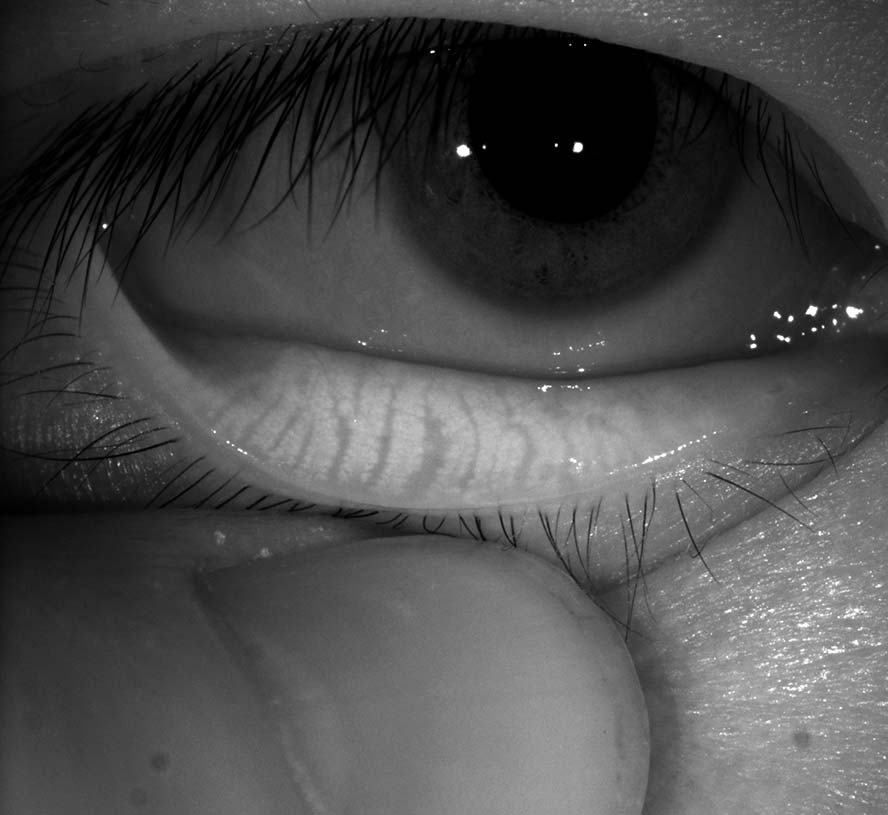
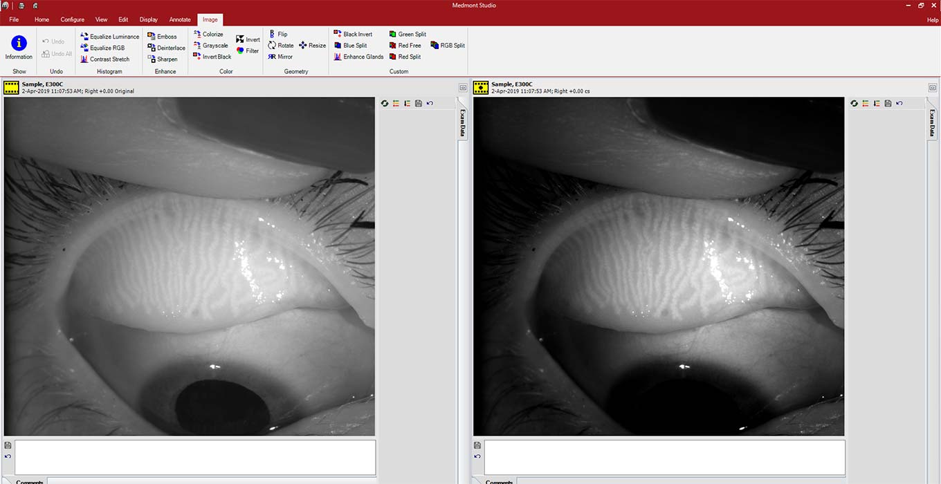
Meibomian Gland Lower
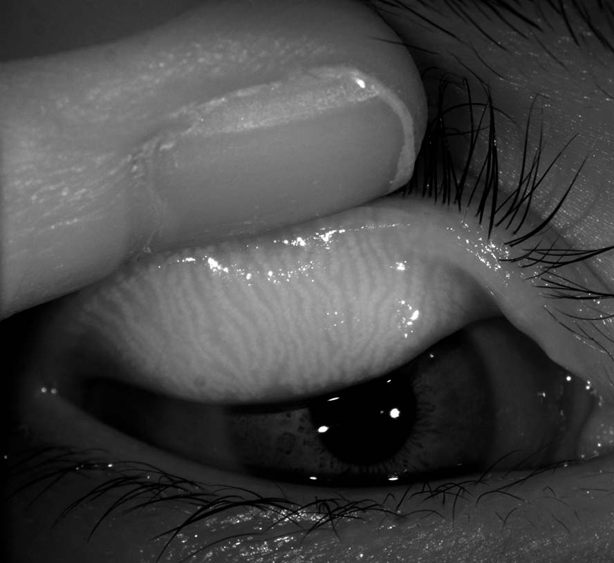
Meibomian Gland Upper
Meibomian Gland Enhanced
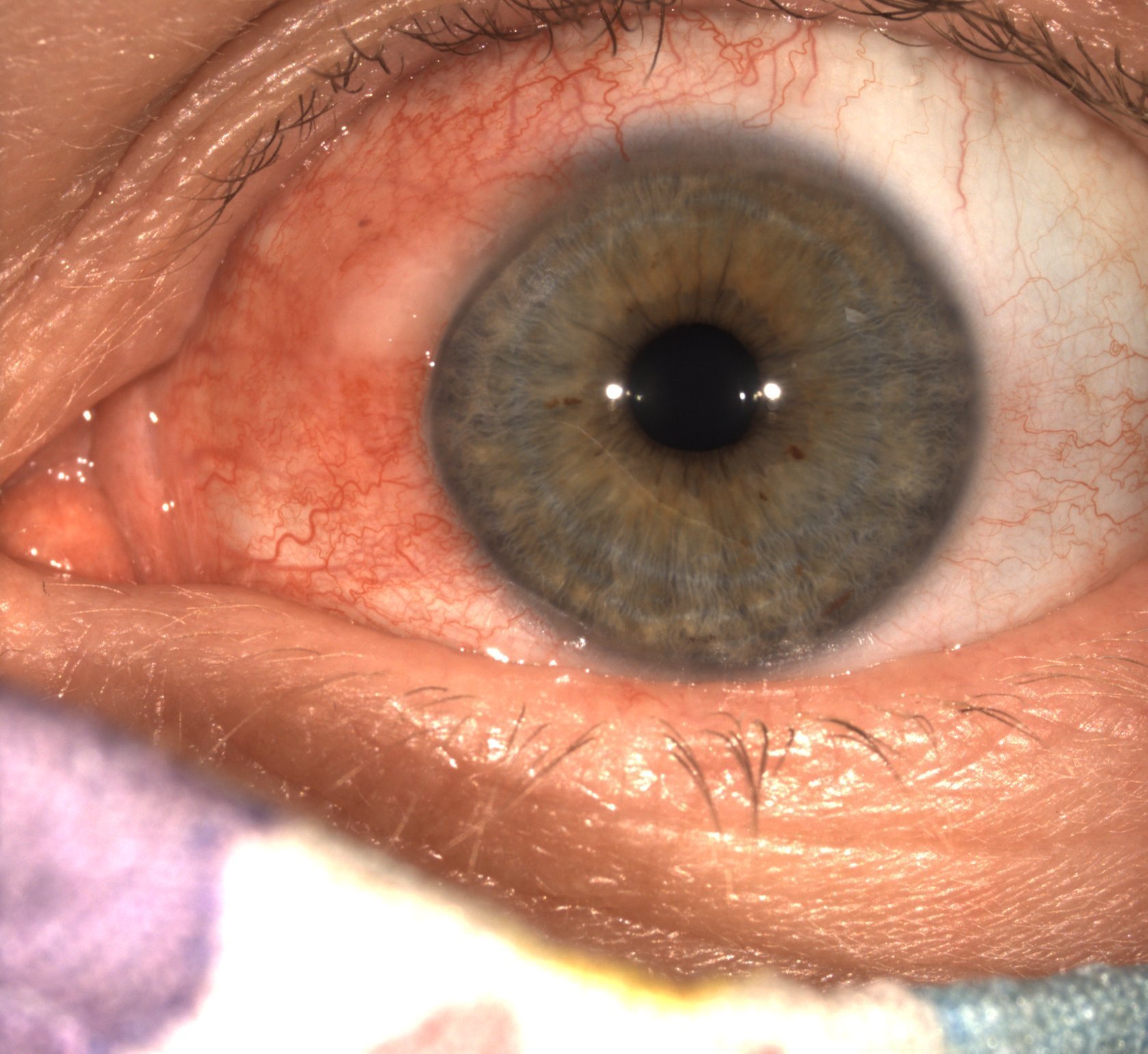
Conjunctival Abrasion
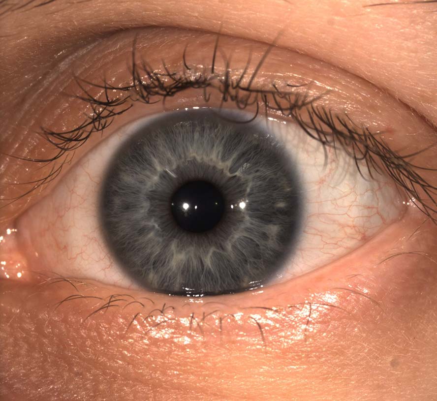
Anterior Image
Scleral Anterior

OrthoK NaFl Video
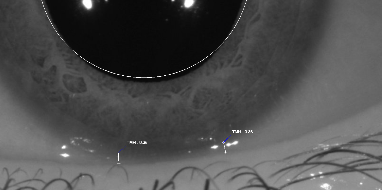
IR TMH Measurement
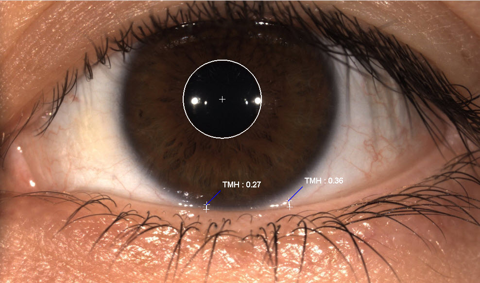
White LED TMH Measurement

Dry Eye – Lissamine green staining

Corneal Staining Incision
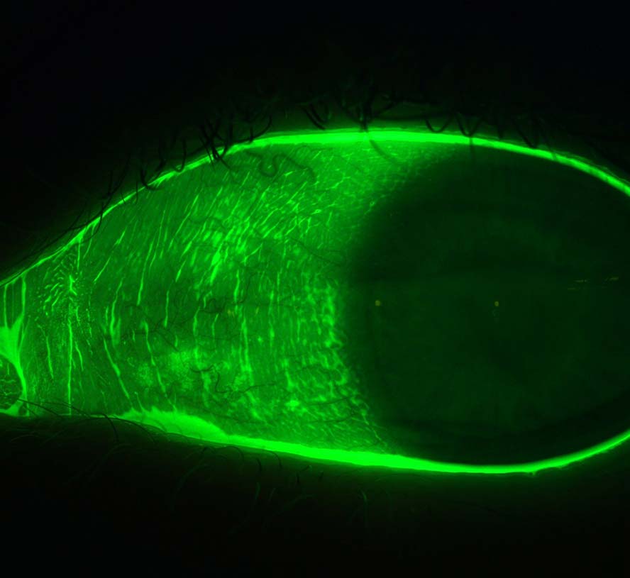
Conjunctival Staining
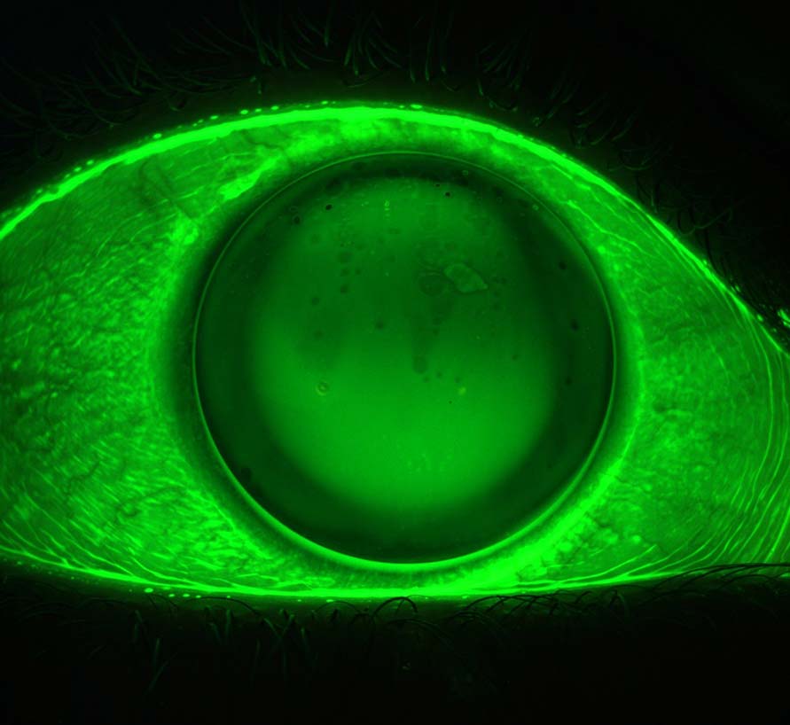
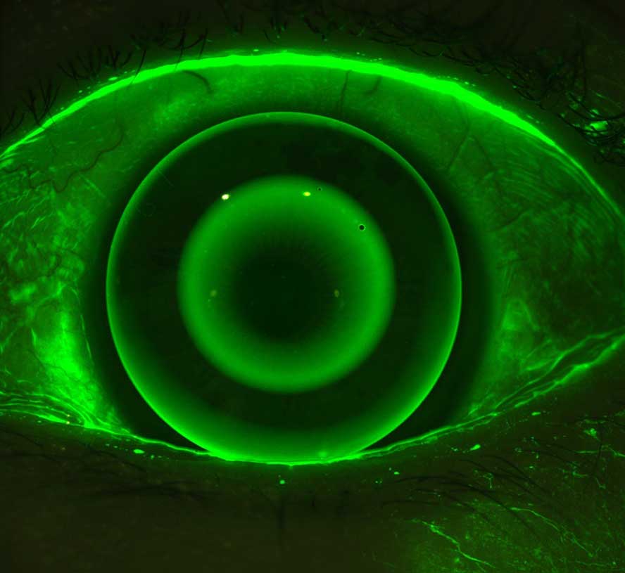
Fluorescien GP Lens
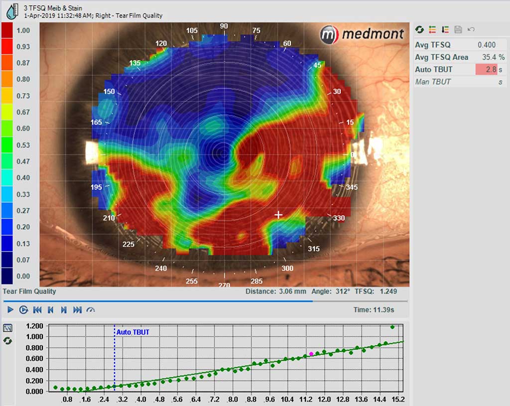
Tear Film Analysis

Non-Wetting
Composite Topography
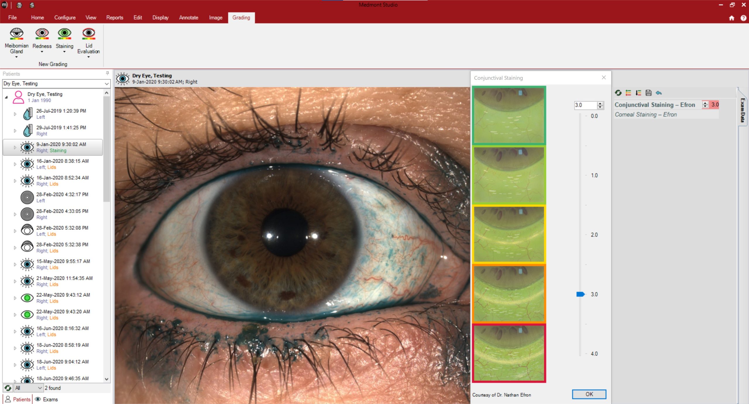
Staining Efron Grading

MGD Efron Grading
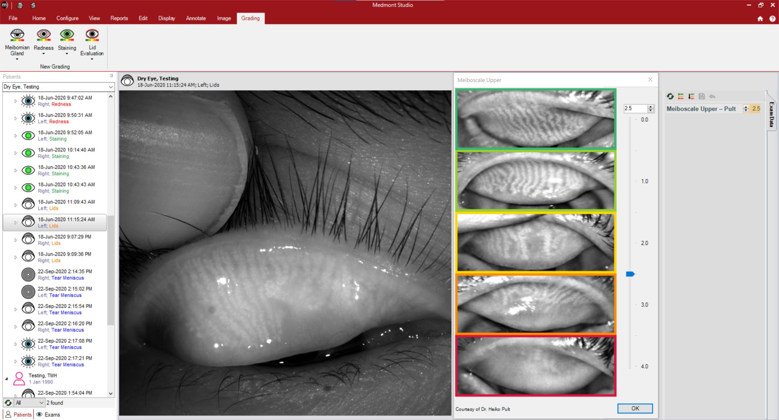
Meiboscale upper grading
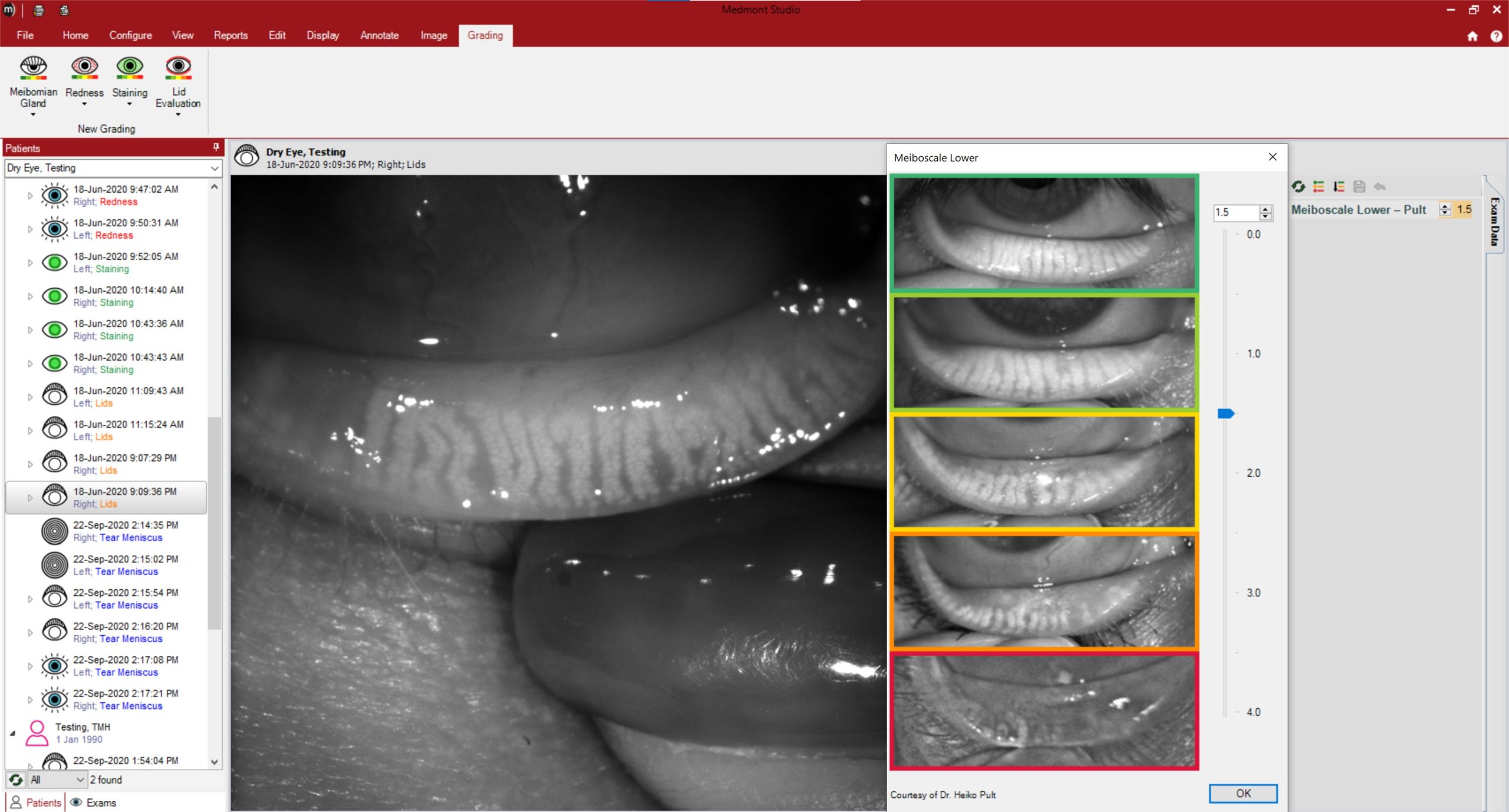
Meiboscale lower grading

Collaboration View
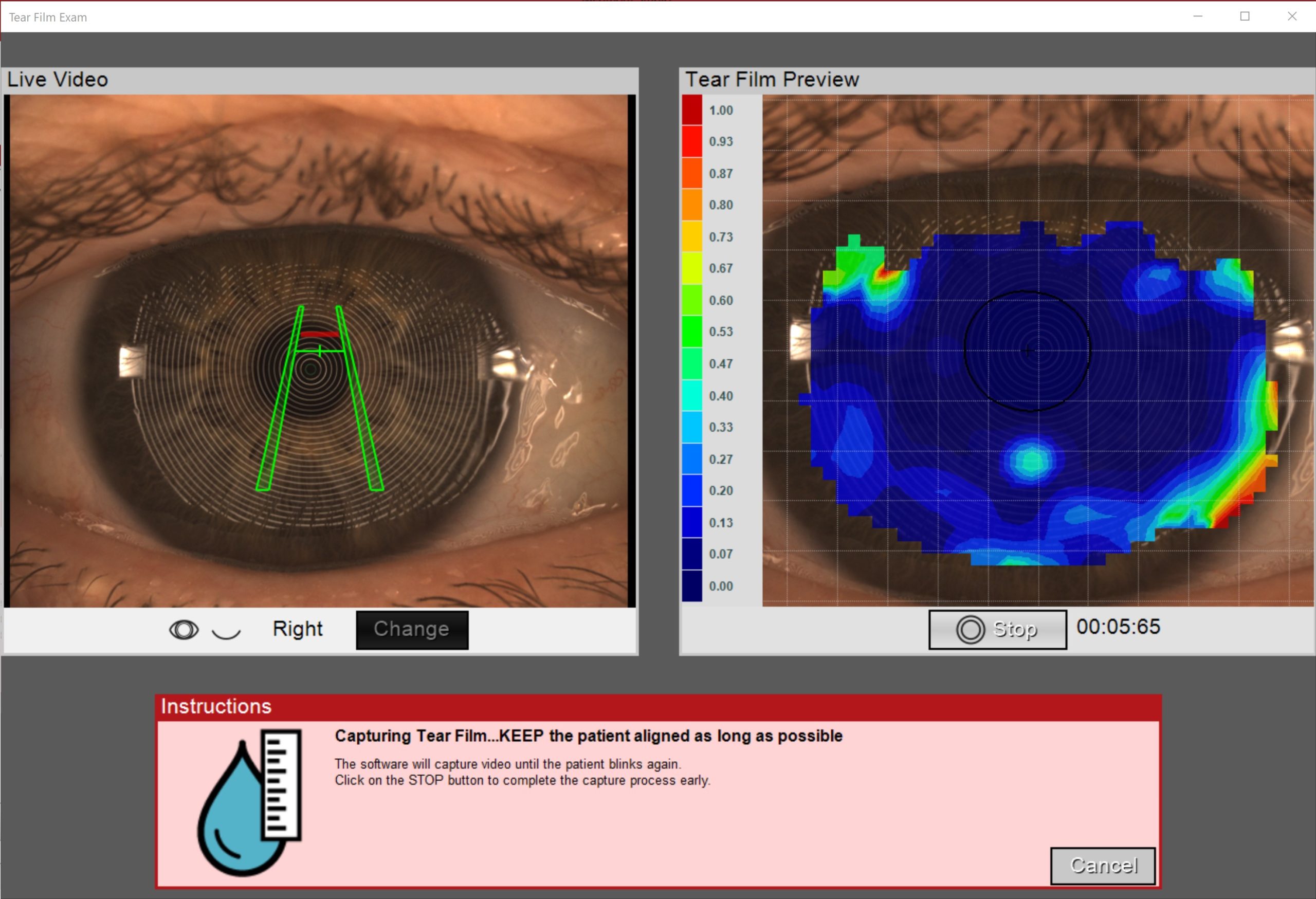
Tear Film Capture 1
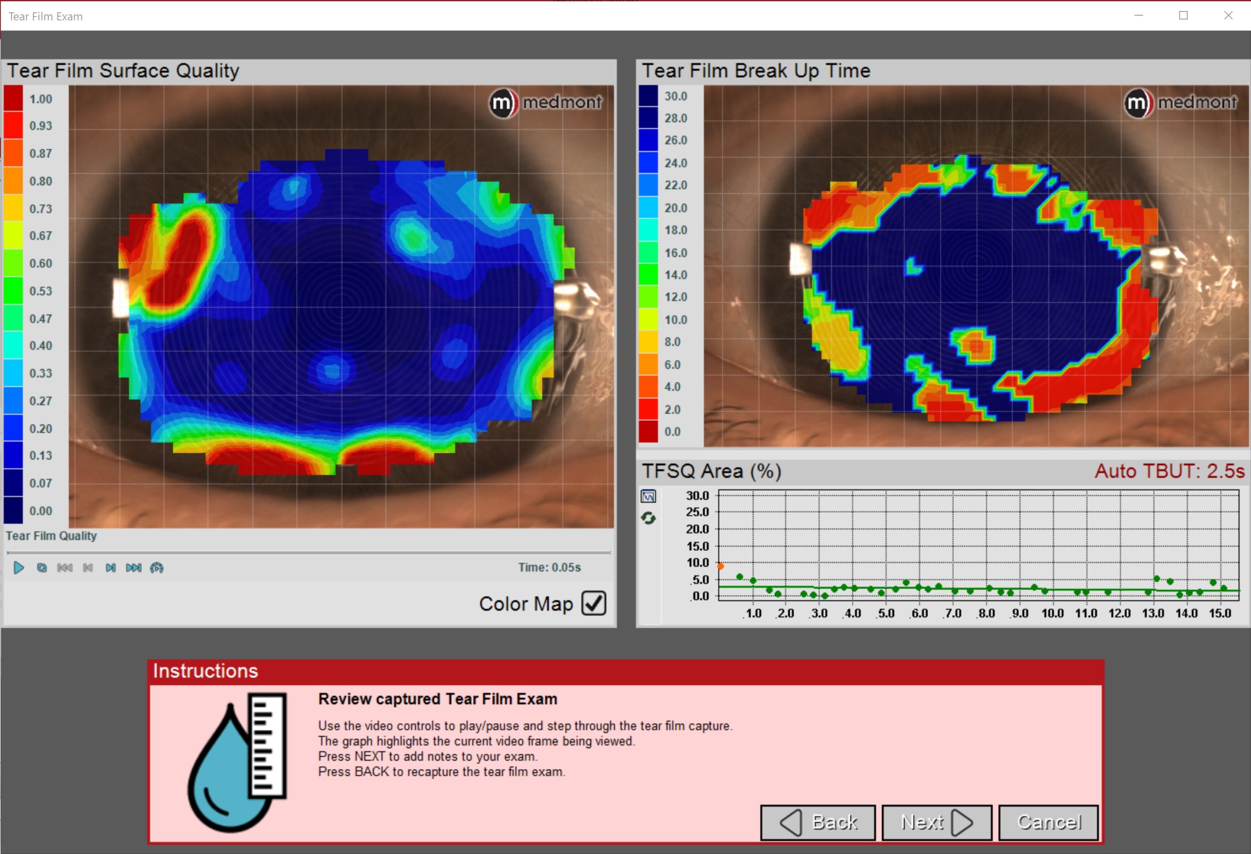
Tear Film Review 1
Anterior Image
Scleral Anterior
Corneal Staining Incision
Conjunctival Staining
Ortho K Fluorescein
Fluorescien GP Lens
Meibomian Gland Upper
Meibomian Gland Lower
Tear Film Analysis
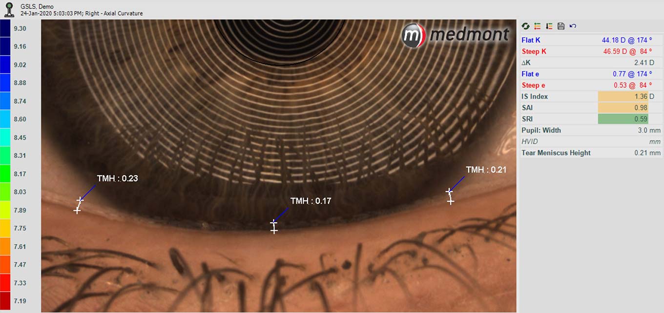
White LED TMH Measurement
Composite Topography
Meibomian Gland Enhanced
WTR Astigmat Composite Topo
Download your free brochure
Fill in the form to tell us where to send your brochure.
Choose between 3 models: Classic, Professional and Vantage
medmont meridia™ Features
Rapid and Precise Computer Aided Image Capture
Superior Performance Through Advanced Image Analysis
Precise Resolution Over Large Area of Coverage
High Capacity Patient Database with Immediate Access to Stored Results Expand Coverage with “Composite Eye” Function
Tear Film Surface Quality Analysis (Still Image & Video)
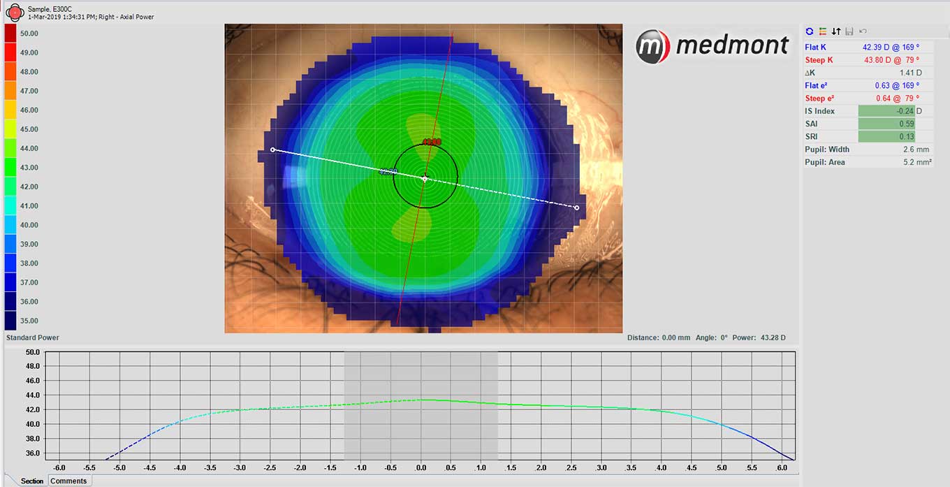
Map Displays:
- Tangential Curvature/Power
- Axial Curvature/Power
- Height
- Elevation from Sphere
- Refractive Power
- Ray Error
- Wavefront Error
- Tear Film Surface Quality Contact Lens Fitting
Contact Lens Fitting:
- Multicurve
- Aspherics
- Keratoconic Designs
- Scleral
- Custom Surfaces
- Custom Laboratory Lens Designs
Shape Descriptors:
- Astigmatism Measurement
- E, p, Q, e2 values
Global Indices:
- SAI
- SRI
- I-S value
Regression Analysis:
- Orthokeratology Subtractive Maps
User Defined Attributes Microsoft Windows™ Based Software::
- Inter/Intra Network Compatible
- EMR/EHR Interface
- DICOM Interface
- USB Computer Interface
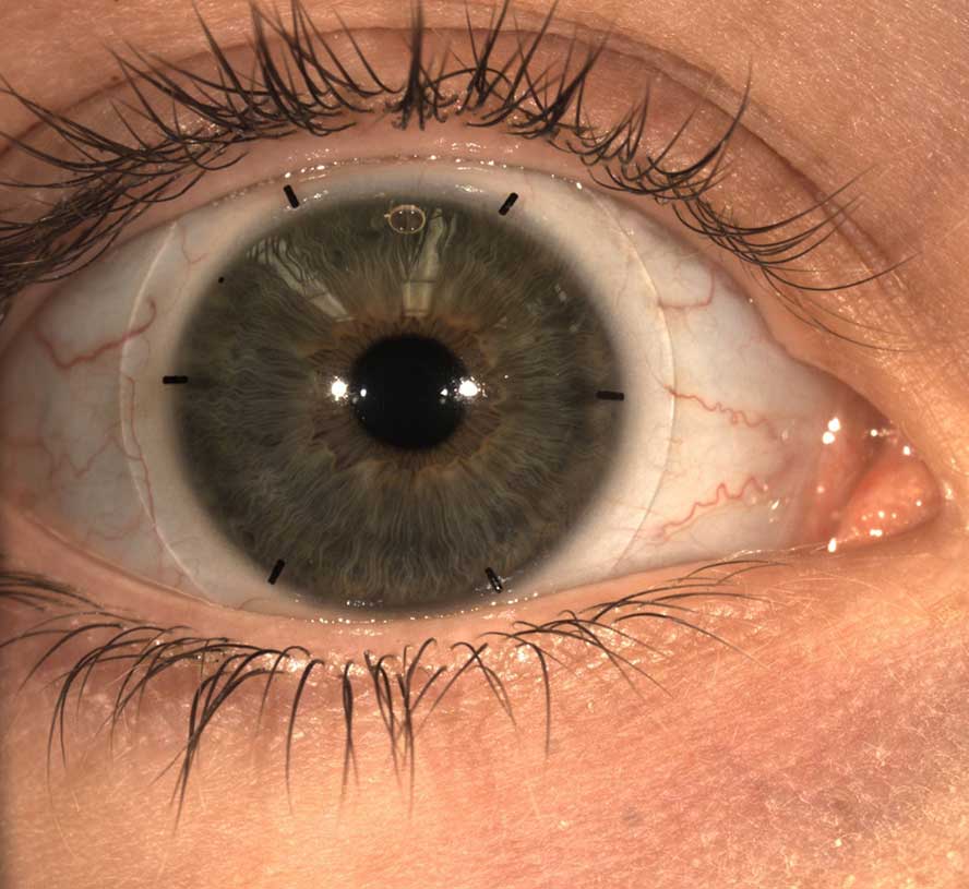
Pupil, Iris, HVID Measurement
Features | Meridia Classic | Meridia Professional | |
Scleral Topography (20mm x 18.5 single capture) | ✓ | ||
Corneal Topography | ✓ | ✓ | ✓ |
Optimized Depth of Field Focus | ✓ | ✓ | ✓ |
High Resolution Digital Color Imaging | ✓ | ✓ | ✓ |
Horizontal Visible Iris Diameter Measurement | ✓ | ✓ | ✓ |
Scleral Lens Simulation | ✓ | ✓ | ✓ |
Ergonomic Quick Keys on Instrument | ✓ | ✓ | ✓ |
Fast and Intuitive Interface Connections | ✓ | ✓ | ✓ |
Anterior Imaging and Video | ✓ | ✓ | |
Meibomian Gland Imaging | ✓ | ✓ | |
Fluorescein Imaging and Video | ✓ | ✓ | |
Focus Guidance Aid | ✓ | ✓ | |
Proven Imaging Grading Scales (EFRON, BHVI, Meiboscale) | ✓ | ✓ | |
Dry Eye Patient Screening Reports | ✓ | ✓ | |
Refined Ergonomics | ✓ | ||
Modern Aesthetics | ✓ | ||
Built-in Calibration Object | ✓ |
Key New Features | |
Increased Field of View Topography | ✓ |
Optimized Depth of Field Focus | ✓ |
High Resolution Digital Color Imaging | ✓ |
Horizontal Visible Iris Diameter Measurement | ✓ |
Scleral Lens Simulation | ✓ |
Medmont Studio 7 Improved User Experience | ✓ |
Ergonomic Quick Keys on Instrument | ✓ |
Convenient and Secure Calibration Ball Storage | ✓ |
Enhanced and Quicker Interface Connections | ✓ |
Anterior Imaging and Video | ✓ |
Meibomian Gland Imaging | ✓ |
Fluorescein Imaging and Video | ✓ |
Tear Meniscus Height Measurements | ✓ |
Scotopic and Photopic Pupil Measurements | ✓ |
Focus Guidance Aid | ✓ |
Imaging Grading Scales | ✓ |
Screening Reports | ✓ |
Key New Features | |
Increased Field of View Topography | ✓ |
Optimized Depth of Field Focus | ✓ |
High Resolution Digital Color Imaging | ✓ |
Horizontal Visible Iris Diameter Measurement | ✓ |
Scleral Lens Simulation | ✓ |
Medmont Studio 7 Improved User Experience | ✓ |
Ergonomic Quick Keys on Instrument | ✓ |
Convenient and Secure Calibration Ball Storage | ✓ |
Enhanced and Quicker Interface Connections | ✓ |
Anterior Imaging and Video | ✓ |
Meibomian Gland Imaging | ✓ |
Fluorescein Imaging and Video | ✓ |
Tear Meniscus Height Measurements | ✓ |
Scotopic and Photopic Pupil Measurements | ✓ |
Focus Guidance Aid | ✓ |
Imaging Grading Scales | ✓ |
Screening Reports | ✓ |
See more. Do more.
Use the 3D/360 product viewer below to explore the Medmont Meridia™.

Specifications
Size
- Weight: 10kg
- Height: 470mm
- Width: 235mm Depth: 345mm
- Shipping Size: 416mm x 416mm x 660mm, 15 kg
Electrical
- Rated Supply Voltage: 100-240 VAC, 50/60 Hz
- Rated Input: 0.19 amps MAX
- Isolation Transformer: Medical Grade, compliant with IEC 60601-1 Min. 500W, min. 4x IEC C13 Outlets, specified for use at national mains voltage
Performance
- Coverage:
Standard: 0.25 –11mm TCC
Limbus to Limbu Data Point Coverage: Limbus to 18mm (composite) - Field of View:
Topography: 17.5 mm (H)
Fluorescein: 20.0 mm (H)
Anterior Image: 26.0 mm(H)
Meibomian Image: 26.0 mm (H) - Repeatability/Accuracy (Test Object): 0.1 Diopters
- Power Range: 10 –100 Diopters
- Number of Rings: 32
- Measurement Points: 9600
Computer Minimum Requirements
- PC and Mains Powered Peripherals: EN/IEC60950 Compliant
- PC Requirements:
- Windows 11 Professional and Home (64 bit) Professional recommended.
- Windows 10 Professional and Home (64 and 32 bit) Professional recommended.
- Windows Server 2016, 2019 and 2022 (64-bit editions only).
- Windows Server “Server Core” installations are not supported.
- Processor: Intel™ i5 generation 6 or later Motherboard: Genuine Intel™ chipset highly recommended.
- Memory: 8 GB for non-video captures, 16 GB for video captures.
- Hard Disk Space: 40 GB for non-video, 200GB for video captures (More for larger databases or busy practices).
- Video cards: GPUs with dedicated memory of 2 Gig or more are recommended.
- Screen resolution: Recommended 1920 x 1080; Minimum supported: 1280 x 720
- USB: USB 3.1 Gen 1 Compliant port on the PC
ALWAYS FOLLOW THE DIRECTIONS FOR USE