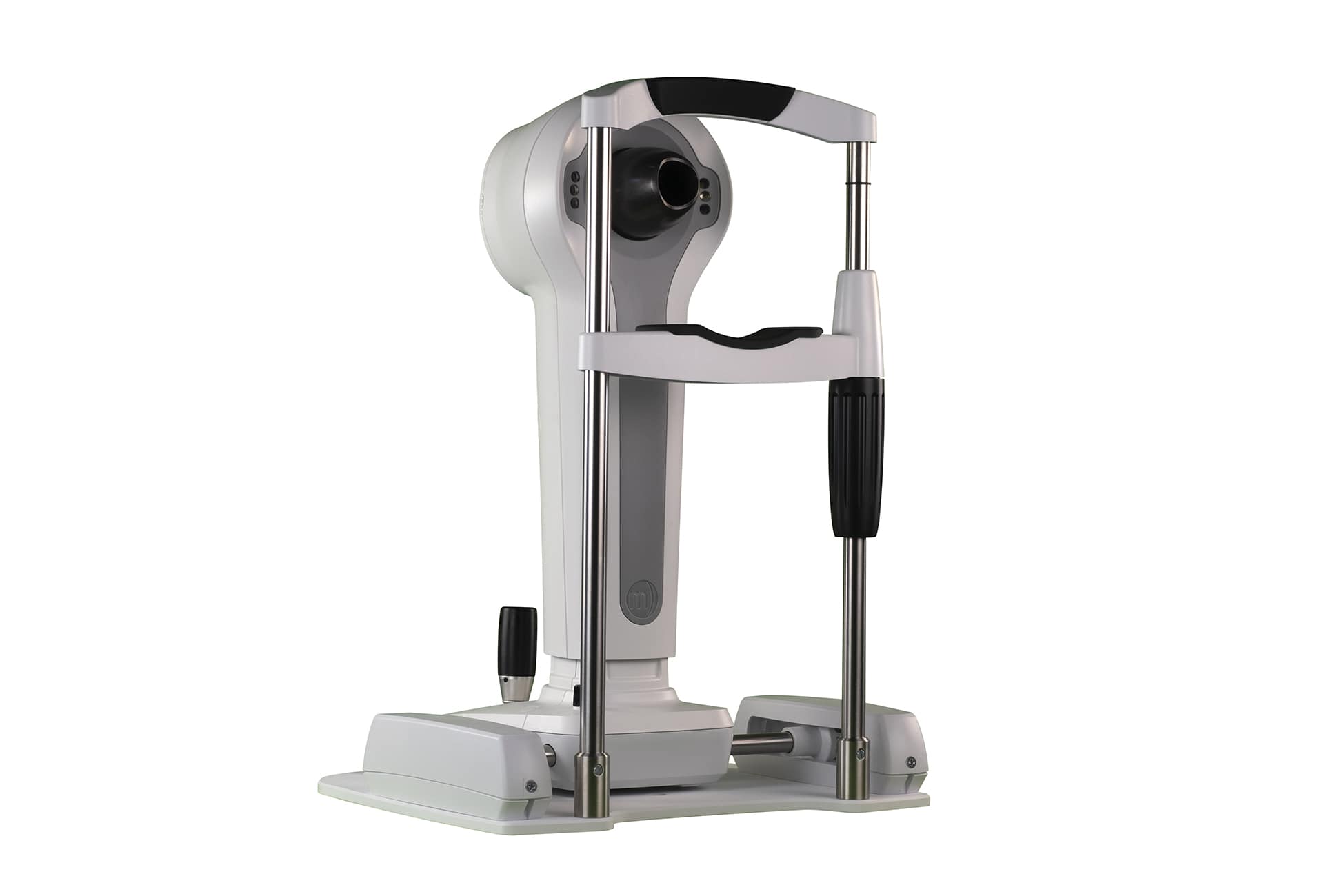
The 5 Ws of Corneal Topography
Corneal topography provides practitioners with a highly desirable map of a patient’s eye.
While corneal topography is standard for many practices, there are still O.D.s who question the value it may provide to their patients. Some O.D.s wonder if adding a topographer will enhance their practice, save precious exam time, or provide a decent return on investment, creating value for their customers and business.
Take a peek at the 5 Ws of corneal topography to help determine if it’s time for your practice to add a new tool to your toolbox (or upgrade your current topographer) to provide your patients an optimal experience.
Who should use corneal topographers to treat patients?
If you have asked whether a corneal topographer is right for your practice, then most likely it is. Topographers have come a long way over the last few decades. They can map scarring from injuries, monitor growths such as pinguecula and pterygium, detect astigmatisms and keratoconus, assist in cornea assessments pre- and post-operatively, and can help diagnosis myopia and subsequently be beneficial in OrthoK lens fittings.
In short, if you seek to provide these options to your patients to specialize your care and avoid the competition of large chain stores and conglomerates, than you are the “who” that should add a corneal topographer to their practice.
What are the differences between placido disc and slit-scanning topographers?
Corneal topography has advanced significantly since its introduction by Father Christopher Scheiner in the 17th century. Today, the most common types of topography are placido disc and slit-scan.
Placido disc topography projects a concentric annular light source on the surface of the cornea and captures reflected light to measure curvature, irregularities, foreign bodies, tear film breakup and other anterior cornea characteristics. While large cone topographers may be considered easier to manipulate, small cones are repeatable and collect more data points therefore are ultimately more precise. Slit-scan is an elevation based method whereby multiple slits are used to assess the corneal surface. The primary difference in slit-scan technology is the output includes posterior cornea data.
When should I implement corneal topography in my exams?
Imaging done with corneal topographer is painless and rarely even slightly uncomfortable. It takes just a few seconds. To get a complete view, though, it may need to be repeated a few times. From start to finish, a topographic scan takes only a few minutes. The scan should be done on non-dilated eyes as the brightness of the instrument may cause glare, making it difficult for the patient to look at the target. This may result in poor mapping. That’s why proper training is invaluable. When you are confident in your abilities to provide corneal topography, your patients feel it and you see it when they return year after year.
Why do you need a corneal topographer?
Are you still unsure of whether a corneal topographer is the right investment for your practice? Consider how easy, comprehensive, and accurate image captures are with the right tool. From assessing and monitoring eye health, including dry eye disease screening, to fitting contact lenses, topographers offer clarity in your approach, convenience in your workflow, and confidence in your decision. Plus, a corneal topography tool elevates the patient experience, differentiates you from large competitors, allows you to develop and expand business and provides your community a full-service, specialized clinic. All of which brings values to your patients and optimizes your ROI!
Where can I get more information and schedule a demo?

See through the fog and find out how to add one tool with many solutions to your optometry practice. Know more and know now when you contact our team to get more information and schedule a demo of our diagnostic systems.
